Gastroenterology Specimens
Gastrointestinal Endoscopy Real Tissue Ex-vivo Models
Check out some of our specimens below. Feel free to reach out for custom specimens.
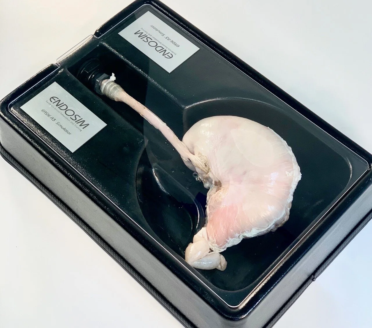
ESD/EMR Porcine Stomach in our EASIE-R3 Simulator
Upper GI Specimen
Porcine ex-vivo esophagus-stomach-duodenum specimen
Diagnostic EGD
Endoscopic Mucosa Resection (EMR)
Endoscopic Submucosal Dissection (ESD)
Variceal band ligation
Barretts Esophagus
Optional Simulated Stenosis -dilation and stent placement
Optional MEGA Specimen- POEM
Bleeding Bovine Colon in our EASIE-R1 Simulator
Lower GI Specimen
Bovine or Porine ex-vivo colon specimen
EMR/ESD in the colon and rectum
Optional Simulated Stenosis -dilation and stent placement
Upper Gastrointestinal Bleeding Specimen in our EASIE-R1 Simulator
Upper/Lower Gastrointestinal Bleeding Specimen
Standard porcine ex-vivo esophagus-stomach-duodenum specimen or Bovine Colon Specimen with 5-6 bleeding vessels sutured into the specimen
Hemostasis in the GI tract:
Injection
Cautery
Clipping
Suturing
Upper / Lower Gastrointestinal Polypectomy Specimen
Artificial polyps in our Upper GI Specimen, or Lower GI Specimens
Upper GI - Standard porcine esophagus, stomach, duodenum with 10-15 polyps
Lower GI Bovine - Standard bovine colon with 10-15 polyps
Lower GI Porcine - Standard porcine colon with 10-15 polyps
* Please contact us for specific types/sizes.
Porcine Stomach in our EASIE-R3 Simulator
Gastric Defect Specimen
Standard Porcine ex-vivo esophagus-stomach-duodenum specimen with EMR type defects
Perforation Closure
Clipping
Suturing
ERCP NeoPapilla Cartridge
ERCP NeoPapilla Cartridge
Cartridge (hybrid) ERCP model with porcine ex-vivo duodenum with an artificial major papilla made of chicken heart tissue.
The artificial papilla allows access to the common bile duct and pancreatic duct. The exchange of the chicken heart allows the performance of multiple sphincterotomies in the same specimen. The chicken heart tissue can be exchanged within 30 seconds.
Diagnostic and therapeutic ERCP
Multiple spincterotomies in the same specimen
15-20 Chicken heart papillae with each specimen cartridge!
Porcine Stomach in our EASIE-R3 Simulator
ERCP Native Biliary
Porcine ex-vivo esophagus-stomach-duodenum-biliary tract-liver-gallbladder specimen.
This complete biliary model contains the native (unaltered) biliary tract including major duodenal papilla, common bile duct, cystic duct, gallbladder, and intrahepatic ducts. Please note that the porcine major papilla does not have a common confluence of common bile duct and pancreatic duct. In the native porcine specimen, the pancreatic duct enters the duodenum at the minor papilla, separate from the common bile duct.
Diagnostic and therapeutic ERCP
One sphincterotomy per specimen
Enteroscopy specimen in our EASIE-R1 Simulator
Balloon Enteroscopy Specimen
Porcine ex-vivo esophagus, stomach, duodenum and 5-6 feet of small bowel (ileum)
Single balloon enteroscopy
Double balloon enteroscopy
Radial EUS image
EUS Complete Specimen
Complete porcine ex-vivo upper GI tract model with esophagus, stomach, duodenum, liver, pancreas, and kidneys. The aorta can be perfused with a roller pump system to simulate Doppler. Optional pathologies on request for additional cost
Diagnostic and therapeutic EUS
EUS-FNA of pancreatic pseudocysts
EUS-FNA of artificial tumors in the esophagus, stomach and liver
EUS-guided cystogastrostomy
Customizable
FNA of Solid lesion in our EUS Task Trainer
EUS Task Trainer
EUS cartridge model with pancreatic cyst, esophageal tumors and gastric mass
Therapeutic EUS
EUS-FNA of target lesions
EUS-FNA of pancreatic pseudocysts
Twisted Sleeve
Porcine ex-vivo esophagus-stomach-duodenum specimen with altered anatomy resembling the status post laparoscopic sleeve gastrectomy
Twisted sleeve model realistically simulates a gastric volvulus as a complication of laparoscopic sleeve gastrectomy
Zenker’s
Cartridge task trainer with ex-vivo model of an artificial pharyngoesophageal diverticulum with a pharyngeal pouch
Zenker diverticulotomy
Roux-en-Y Gastric Bypass
Porcine ex-vivo esophagus-stomach-duodenum specimen with altered anatomy resembling the status post laparoscopic Roux-en-Y gastric bypass.
This model has the NeoPapilla attachment that allows sphincterotomy and therapeutic ERCP.
EGD and ERCP in Roux-en-Y Gastric Bypass Anatomy
Proctoscopy
Cartridge model of a porcine ex-vivo anus and rectum
Proctoscopy
Hemorrhoid ligation
Anal sphincter repair


